Key Takeaways
- ECG and EKG measure your heart’s electrical activity to assess rhythm and rate.
- The test helps detect heart problems like arrhythmias or signs of past heart damage.
- Knowing how the test works prepares you to discuss results confidently with your doctor.
When your heart beats, it sends tiny electrical signals through your body. An electrocardiogram—known as an ECG or EKG—records those signals to show how your heart is working. This simple, painless test reveals how fast your heart beats, how steady its rhythm is, and whether there are signs of heart strain or disease.
You might have an ECG during a routine checkup, before surgery, or if you experience chest pain, dizziness, or shortness of breath. The test helps your healthcare provider spot irregular heartbeats, past heart attacks, or changes that suggest blocked arteries. In just a few minutes, it can uncover details about your heart’s health that you can’t feel but matter deeply.
Understanding how ECG and EKG tests work gives you more control over your heart health. Knowing what to expect—and what the results mean—helps you take informed steps toward protecting your heart for the long run.
Understanding ECG and EKG
An electrocardiogram records the heart’s electrical activity to show how well it functions. You can use it to identify irregular rhythms, signs of heart strain, or evidence of past or current heart problems. Both ECG and EKG describe the same test that helps clinicians assess your heart’s electrical patterns safely and quickly.

What Is an Electrocardiogram?
An electrocardiogram (ECG or EKG) measures the tiny electrical impulses that make your heart beat. Each impulse travels through specific pathways, causing the heart’s chambers to contract and pump blood.
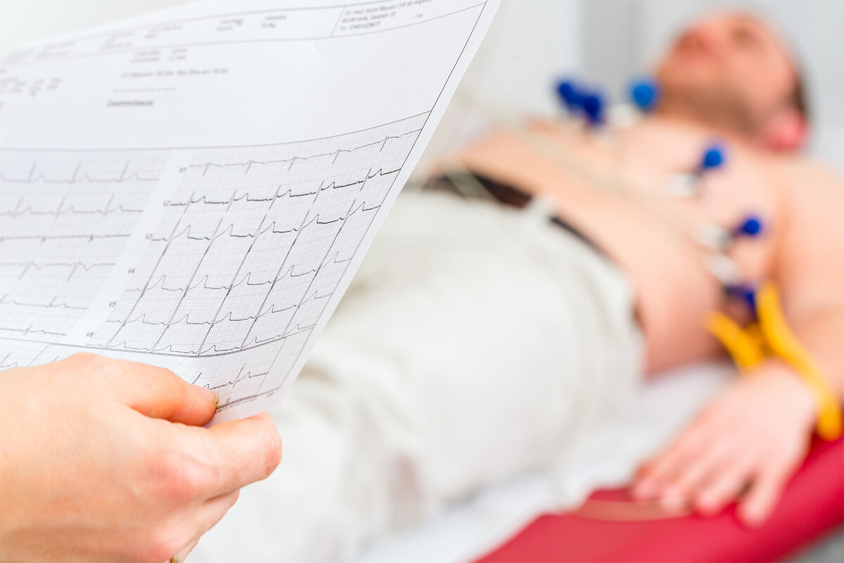
During the test, a healthcare professional places small electrodes on your chest, arms, and legs. These sensors detect electrical activity and send it to a monitor or printout. The results appear as waves that represent each heartbeat’s timing and strength.
You don’t need special preparation for a standard ECG. The procedure is painless and takes only a few minutes. Some tests, such as an exercise ECG or Holter monitor, record your heart’s activity during physical movement or over a longer period to detect rhythm changes that occur outside a clinical setting.
Difference Between ECG and EKG
ECG and EKG refer to the same diagnostic test. The difference lies only in the abbreviation. “ECG” comes from the English electrocardiogram, while “EKG” originates from the German elektrokardiogramm.
Medical staff in some regions prefer “EKG” to avoid confusion with EEG, which measures brain activity. Regardless of spelling, both terms describe the same process of recording your heart’s electrical signals.
| Term: ECG | |
|---|---|
| Language Origin | English |
| Meaning | Electrocardiogram |
| Term: EKG | |
|---|---|
| Language Origin | German |
| Meaning | Elektrokardiogramm |
Understanding that these terms are interchangeable helps you interpret medical forms and discussions more easily. You can use either abbreviation when speaking with your healthcare provider.
Why These Tests Matter
An ECG or EKG helps detect a range of heart issues, including arrhythmias, heart attacks, and electrolyte imbalances. It can also reveal whether your heart’s chambers are enlarged or under strain.
Doctors often use this test when you report chest pain, shortness of breath, or irregular heartbeats. It can also serve as a routine screening if you have risk factors such as high blood pressure or diabetes.
Because the test is quick, non-invasive, and inexpensive, it’s one of the most common tools for evaluating heart health. Your provider can review the results immediately and decide whether further testing or treatment is needed.
How ECG and EKG Tests Work
An ECG or EKG measures the electrical activity that keeps your heart beating in rhythm. It uses small sensors, a recording machine, and various test types to capture how your heart functions during rest or activity.
The Role of Electrodes
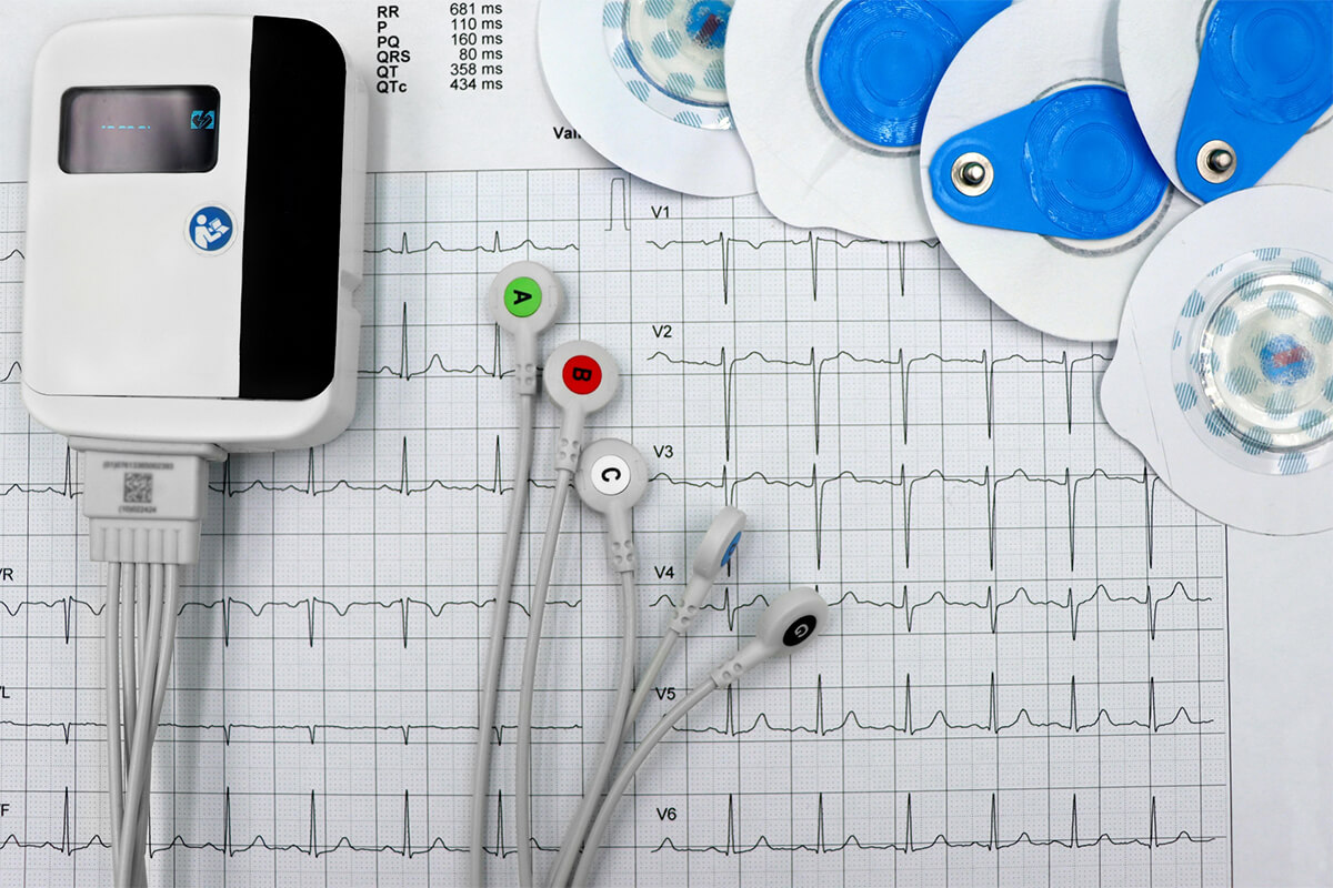
Electrodes are small, sticky patches placed on your chest, arms, and legs. They detect the electrical signals that move through your heart with each beat. These signals travel through thin wires to the EKG machine for recording.
Technicians may clean or shave small areas of skin to help the electrodes stick better. You can breathe normally during the test, but staying still helps prevent interference.
Each electrode plays a role in mapping your heart’s electrical pattern. Together, they create a complete picture of how efficiently your heart’s chambers contract and relax.
| Placement Area: Chest | |
|---|---|
| Purpose | Tracks main heart activity |
| Placement Area: Arms and Legs | |
|---|---|
| Purpose | Provides reference signals for comparison |
The EKG Machine and How It Records
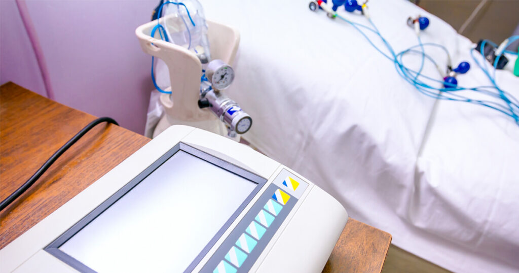
The EKG machine receives signals from the electrodes and converts them into a visual trace called a waveform. Each wave represents a phase of your heartbeat — the P wave, QRS complex, and T wave show how electrical impulses travel through the heart.
You usually see these waves printed on paper or displayed on a monitor. The machine measures timing and strength, allowing your healthcare provider to identify irregular rhythms or signs of previous heart damage.
Modern EKG machines can store data digitally and compare results over time. Portable versions, including smartwatch sensors, can record limited single-lead readings for personal monitoring.
Types of ECG/EKG Tests
Different test types help doctors understand how your heart behaves in various conditions.
- Resting ECG: Done while you lie still. It provides a baseline of your heart’s normal rhythm.
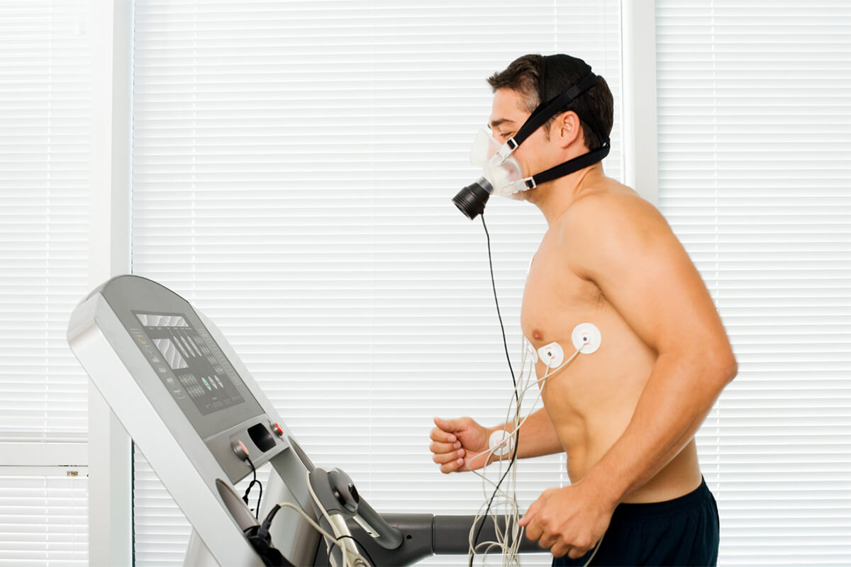
- Stress ECG: Performed while you walk or run on a treadmill. It shows how your heart responds to exertion.
- Holter Monitor: A small device you wear for 24–48 hours to record continuous heart activity.
- Event Monitor: Used for several weeks. You activate it when you notice symptoms like palpitations.
Each test offers unique insights. Your provider chooses the one that best matches your symptoms and medical history.
What ECG and EKG Reveal About Heart Health
An electrocardiogram shows how your heart’s electrical activity works in real time. It helps your healthcare provider understand how your heart beats, identify rhythm changes, and detect signs of damage or reduced blood flow.

Heart Rate and Rhythm Insights
An ECG or EKG measures how fast and how regularly your heart beats. The test records the electrical signals that trigger each heartbeat, producing a visual pattern of waves.
Your heart rate—the number of beats per minute—shows whether your heart is working within a healthy range. A normal resting rate for most adults is 60–100 beats per minute.
The heart rhythm tells whether your heartbeat follows a steady pattern. Regular spacing between waves suggests a stable rhythm, while uneven spacing may indicate an irregularity.
Doctors review these patterns to see if your heart beats too fast (tachycardia), too slow (bradycardia), or irregularly. Even if you feel fine, subtle changes in rhythm can point to early heart issues or the effects of medications.
Detecting Arrhythmias
An arrhythmia occurs when the heart’s electrical impulses misfire, causing irregular beats. ECGs detect these changes by capturing the timing and shape of each electrical signal.
Common arrhythmias include:
- Atrial fibrillation (AFib): rapid, uneven beating in the upper chambers.
- Atrial flutter: similar to AFib but with a more organized pattern.
- Ventricular tachycardia: fast rhythm from the lower chambers.
You might not always feel symptoms, but an ECG can reveal skipped beats, pauses, or extra beats that you don’t notice. Detecting arrhythmias early helps prevent complications such as stroke or heart failure.
If your doctor suspects an intermittent rhythm problem, you may wear a Holter monitor or event monitor at home. These devices record your heart’s activity over time to catch irregularities that a short test might miss.
Identifying Heart Attack Signs
An ECG can show whether your heart has experienced or is currently experiencing a heart attack. It does this by detecting changes in the wave patterns that reflect how electrical signals move through heart muscle tissue.
When part of the heart lacks oxygen due to a blocked artery, the electrical pattern shifts. Certain segments of the ECG trace—such as the ST segment—may rise or fall, signaling reduced blood flow or tissue injury.
Doctors also use ECG results to determine which part of the heart is affected and how severe the damage may be. Even after recovery, the test can show evidence of a previous heart attack.
If you have chest pain, shortness of breath, or dizziness, an ECG helps your healthcare team quickly assess whether your symptoms relate to heart muscle damage or another cause.
What Your Doctor Looks For in Your ECG Results
Your ECG reveals how your heart’s electrical system functions with each beat. It shows whether your heart rhythm, rate, and electrical signals follow normal patterns or suggest a possible issue that needs more attention.
The Rhythm of Your Heartbeat (Is it steady or irregular?)
Your doctor studies the rhythm strip on your ECG to see if your heartbeats occur at regular intervals. A steady rhythm means the electrical impulses travel smoothly through the heart’s conduction system. Irregular rhythms, such as atrial fibrillation or premature beats, may show inconsistent spacing between the peaks on the tracing.
The P wave is a key part of this check. It represents the electrical activity that starts in the atria. If the P waves appear uneven or missing, it can point to irregular atrial activity or an abnormal pacemaker focus.
Doctors often use a table like this to interpret rhythm findings:
| Pattern: Regular spacing | |
|---|---|
| Possible Meaning | Normal sinus rhythm |
| Pattern: Irregular spacing | |
|---|---|
| Possible Meaning | Atrial fibrillation or ectopic beats |
| Pattern: Missing P waves | |
|---|---|
| Possible Meaning | Junctional or ventricular rhythm |
Recognizing these patterns helps your doctor decide whether your heart is beating in a coordinated way.
The Speed of Your Heartbeat (Is it too fast or too slow?)
Your heart rate is calculated by measuring the distance between R waves in the QRS complex. This tells your doctor whether your heart beats within a normal range—usually 60 to 100 beats per minute for adults at rest.
A rate above 100 may indicate tachycardia, while below 60 may suggest bradycardia. These findings help determine if your heart’s natural pacemaker, the sinoatrial node, is functioning properly.
A short list of rate interpretations:
- Normal rate (60–100 bpm): Healthy sinus rhythm
- Too fast (>100 bpm): Possible stress, fever, or arrhythmia
- Too slow (<60 bpm): Could reflect athletic conditioning or conduction delay
Your doctor uses this information along with your symptoms to decide if further tests or treatments are needed.
The Strength and Timing of Electrical Signals
Each ECG wave shows how electrical signals move through your heart. The P wave, QRS complex, and T wave must appear in a specific order and shape for the heart to contract efficiently.
The QRS complex represents the ventricles contracting. If it’s too wide or oddly shaped, that can mean the signal is delayed or blocked. The T wave follows, showing the ventricles resetting for the next beat. Abnormal T waves may suggest electrolyte imbalances or signs of strain on the heart muscle.
Doctors also measure intervals, such as the PR interval and QT interval, to assess timing. Longer or shorter intervals can reveal conduction issues or medication effects.
By analyzing these details, your doctor can identify how well your heart’s electrical system coordinates each beat and whether the signals are strong and timely.
Getting Your ECG: What to Expect
An electrocardiogram records your heart’s electrical activity in a few minutes and helps identify rhythm problems or signs of heart disease. The process is simple, safe, and usually done in a clinic, hospital, or even an ambulance.
Preparing for the Test
You don’t need to fast or change your routine before an ECG. Still, tell your healthcare provider about all medications and supplements you take, since some can affect heart rhythm.
Wear loose, comfortable clothing that allows easy access to your chest, arms, and legs. You may be asked to remove jewelry or metal objects that could interfere with the electrodes.
If you have chest hair, a small area might be shaved to help the sensors stick better. Avoid using lotions, oils, or powders on your skin before the test—they can prevent proper contact.
During a stress test, which is a type of ECG done while you exercise, you might need to wear athletic shoes and clothes suitable for walking or running on a treadmill.
Risks and Skin Irritation
An ECG is noninvasive and safe. The sensors, called electrodes, only record electrical signals; they don’t send any current into your body.
Some people notice mild skin irritation where patches are placed. This can look like redness or a small rash, especially if you have sensitive skin. The feeling when removing the adhesive pads is similar to peeling off a bandage.
If discomfort lasts after the test, gently wash the area with mild soap and water. Applying an unscented moisturizer can soothe the skin. In rare cases, an allergic reaction to the adhesive may occur—let your provider know if you’ve had issues with medical tapes or patches before.
What Happens After Your Test
You can usually return to normal activities immediately after an ECG. The test itself takes only a few minutes, and there’s no recovery time needed.
Your results may be available right away if a clinician reads the tracing on-site. Otherwise, your doctor will review the data and contact you later.
If your ECG was part of a stress test, you might rest briefly afterward while your care team monitors your heart rate and blood pressure until they return to normal.
Bring a list of your recent symptoms or episodes of chest pain, dizziness, or palpitations. This helps your provider interpret the results more accurately.
Follow-Up with a Cardiologist
A cardiologist reviews your ECG to look for patterns that show how your heart is working. They check your heart rate, rhythm, and electrical conduction for signs of arrhythmia, heart attack, or poor blood flow.
If the results show irregularities, your cardiologist may recommend additional tests such as an echocardiogram or a stress ECG to assess how your heart performs under exertion.
You might also discuss lifestyle factors like diet, exercise, and stress management. These conversations help your doctor create a plan tailored to your heart health.
Keep a copy of your ECG report for future visits. Comparing results over time helps track changes and guides treatment decisions.